Kidney Ultrasound: What It Shows and How It's Done
AI Summary
Kidney ultrasound is a painless, radiation-free diagnostic method that images kidney structure using sound waves. Used to detect stones, cysts, tumors and obstructions.
What is a Kidney Ultrasound?
Kidney ultrasound (renal ultrasonography) is a diagnostic method that uses high-frequency sound waves to image the structure, size, and potential diseases of the kidneys. It is radiation-free, painless, and safe for all age groups, including pregnant women and children.
When is Kidney Ultrasound Recommended?
- Flank and back pain
- Blood in urine (hematuria)
- Recurrent urinary tract infections
- Suspected kidney stones
- Abnormal kidney function tests
- Palpable abdominal mass
- Investigation of hypertension causes
- Follow-up of kidney cysts or tumors
What Can Kidney Ultrasound Detect?
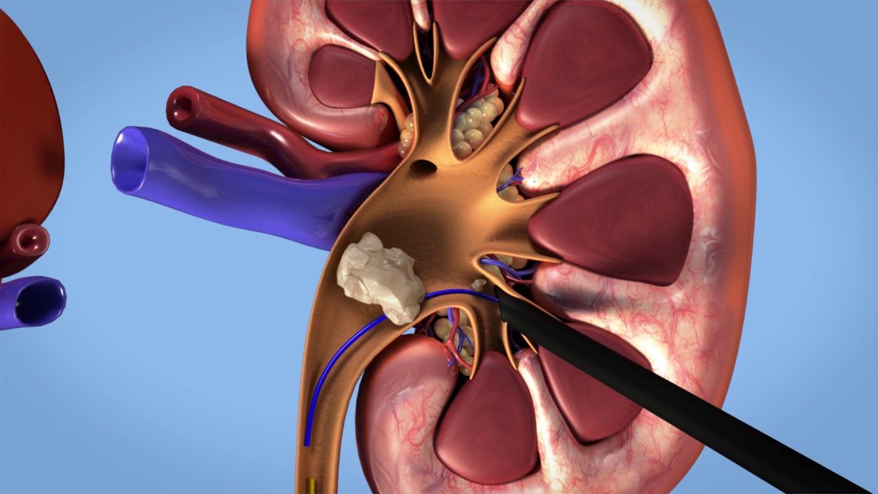
- Kidney stones: Size, number, and location
- Hydronephrosis: Kidney swelling due to urinary obstruction
- Kidney cysts: Simple vs. complex cyst differentiation
- Kidney tumors: Solid masses and suspicious lesions
- Size anomalies: Small or enlarged kidneys
- Congenital anomalies: Horseshoe kidney, duplex collecting system
How is Kidney Ultrasound Performed?
- No special preparation usually required
- Patient lies on back or side
- Gel applied to skin, ultrasound probe placed
- Duration: 10-20 minutes
- Results displayed and interpreted in real-time
Kidney Ultrasound in Izmir
Prof. Dr. Orçun Çelik performs comprehensive urological evaluations including kidney ultrasound during consultations at Izmir Bazekol Hospital.